
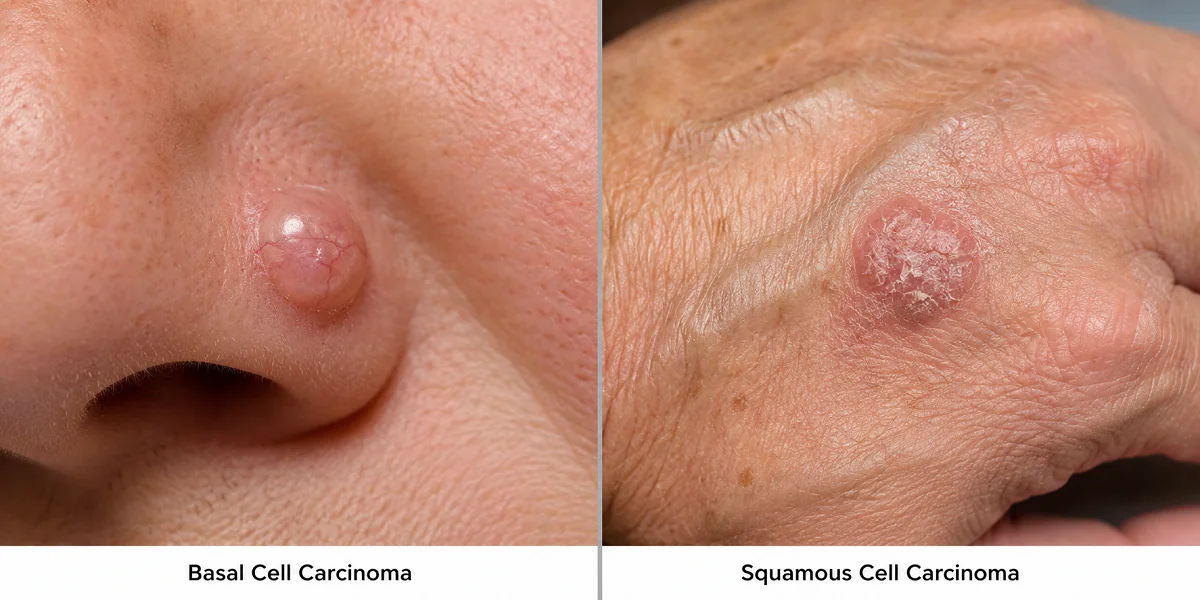
If a dermatologist has just told you that you have a basal cell carcinoma or a squamous cell carcinoma, or you’ve spotted something on your skin that doesn’t look right, you probably want a straight answer to one question: are these the same thing, and does it matter which one it is? It does. Basal cell carcinoma vs squamous cell carcinoma is one of the most common comparisons in dermatology, because these two cancers are frequently mentioned in the same breath. They’re also different enough that mixing them up can lead to the wrong level of concern in either direction.
Here’s the short version. Basal cell carcinoma (BCC) is the more common of the two, grows slowly, and almost never spreads. Squamous cell carcinoma (SCC) is less common, tends to grow faster, and carries a real, though still generally low, risk of spreading if it’s left alone too long. Neither is something to shrug off. Neither is a reason to panic before you’ve even seen a doctor.
What Basal Cell Carcinoma and Squamous Cell Carcinoma Actually Are
Both cancers start in the epidermis, the outermost layer of your skin, but they come from different cells within it. That distinction is the entire reason they’re named differently and behave differently.
Basal cells sit at the bottom of the epidermis. Their job is to keep dividing and pushing new cells upward, replacing older cells as they wear away. When something damages the DNA in these basal cells, usually years of cumulative ultraviolet exposure, they can start growing uncontrolled. That’s basal cell carcinoma.
Squamous cells are the flatter cells higher up in the epidermis, closer to the surface. They’re essentially the basal cells’ descendants, having moved upward and changed shape over their life cycle. When squamous cells mutate and grow out of control instead of shedding normally, that’s squamous cell carcinoma.
Both are grouped under the umbrella term non-melanoma skin cancer, distinguishing them from melanoma, which starts in a completely different cell type (melanocytes) and behaves far more aggressively. It’s a genuinely useful category, but it can also blur the line between BCC and SCC for people trying to understand their own diagnosis. They’re related. They’re not interchangeable.
How Common Is Each One
This part surprises a lot of people. According to the American Cancer Society, about 8 out of 10 skin cancers are basal cell carcinomas, making it the single most common cancer in the United States, skin or otherwise. Squamous cell carcinoma accounts for roughly 2 out of 10 skin cancers, which still adds up to well over a million diagnoses a year. So if you’re comparing raw odds, you’re more likely to be looking at a BCC. That doesn’t mean an SCC diagnosis is rare or unusual. It’s just less frequent than its counterpart.
How They Look Different on Skin
This is usually the part people care about most, and it’s also where the two cancers diverge the most visibly.
Basal cell carcinoma tends to show up as:
- A pearly or waxy bump, sometimes with tiny visible blood vessels running through it
- A flat, slightly scar-like patch that’s flesh-colored or pink
- A sore that bleeds, scabs over, and then reopens in a cycle that doesn’t fully heal
Squamous cell carcinoma tends to show up as:
- A firm, red, raised bump or nodule
- A rough, scaly patch that can look like a stubborn dry-skin spot
- A sore or wart-like growth that crusts, and often bleeds more readily than a BCC would
Neither list is exhaustive, and skin cancer doesn’t always follow the textbook. I’ve seen patients describe what turned out to be a BCC as “just a pimple that wouldn’t go away,” which is a fair way to describe it before you know what you’re looking at. If a spot on your skin has been present for more than a few weeks and isn’t healing normally, that’s the signal to get it looked at, regardless of which list it seems to match.
Growth Rate and Risk of Spreading
This is where the two conditions genuinely part ways in terms of how seriously they’re treated.
Basal cell carcinoma is slow. It can sit on the skin for months, sometimes years, growing gradually enough that people don’t notice until it’s fairly established. The tradeoff is that it almost never metastasizes, meaning it almost never spreads to lymph nodes or distant organs. Left untreated long enough, it can still grow into nearby tissue, including bone in severe cases, so “slow” doesn’t mean “safe to ignore.” It means there’s usually more time to catch it before it causes serious local damage.
Squamous cell carcinoma moves faster and carries a higher, though still relatively low, risk of spreading beyond the skin if it’s not treated. This risk climbs meaningfully in certain situations: lesions on the lips or ears, cancers that develop in old scars or chronic wounds, and cases in patients who are immunosuppressed, such as organ transplant recipients. A clinical review published in American Family Physician found that organ transplant recipients face roughly a 65-fold increased risk of squamous cell carcinoma compared with the general population, which gives you a sense of how much immune status affects this particular cancer’s behavior.
There’s also a well-documented precursor relationship worth knowing about. The same American Family Physician review notes that actinic keratosis, those rough, sandpaper-like patches many people develop after decades of sun exposure, is considered the same disease process as squamous cell carcinoma at an earlier stage. Not every actinic keratosis turns into SCC, but it’s the reason dermatologists take those spots seriously even when they look minor.
Where Each One Tends to Show Up
Both cancers favor sun-exposed skin, which is exactly what you’d expect given that UV exposure is the dominant risk factor for both. Face, ears, neck, scalp, and forearms are the usual suspects.
Basal cell carcinoma leans heavily toward areas that get consistent, cumulative sun exposure over a lifetime, like the nose, cheeks, and hairline. Squamous cell carcinoma shows a similar pattern but is somewhat more likely to appear on the lips, the rims of the ears, and the backs of the hands. It’s also more likely than BCC to develop in skin that’s already damaged in some other way, like a long-standing scar, a burn site, or an area of chronic inflammation.
Diagnosis and Treatment
Here’s something that might be reassuring: the path to diagnosis is essentially identical for both. A dermatologist examines the lesion and, if it looks suspicious, takes a biopsy, a small sample of tissue, to confirm what’s actually going on under a microscope. Visual inspection alone isn’t considered reliable enough for a diagnosis, no matter how experienced the examiner is. That biopsy is what separates a confirmed BCC from a confirmed SCC, and from other conditions that can mimic either one.
Treatment overlaps a lot too. Depending on the size, location, and depth of the lesion, options include:
- Surgical excision, removing the growth along with a margin of healthy tissue
- Mohs micrographic surgery, a precise, layer-by-layer removal technique often used on the face or in cases where the cancer has a higher chance of recurring
- Topical treatments, such as imiquimod or 5-fluorouracil, generally reserved for superficial cases
- Radiation therapy, typically an option when surgery isn’t practical
The main difference isn’t which treatments are available. It’s how urgently treatment gets scheduled and how closely the case gets monitored afterward. Because SCC carries a higher baseline risk of spreading, especially in high-risk locations or high-risk patients, dermatologists tend to move faster and follow up more closely than they might for a straightforward, low-risk BCC.
I’ll be upfront about something here: I’m not a dermatologist, and nothing in this article is a substitute for an actual clinical exam. What I can tell you, based on the research behind this piece, is that both conditions are highly treatable when caught early, and the specific plan for either one really does depend on details a biopsy and an exam will reveal that no article can.
When to See a Dermatologist
You don’t need to correctly diagnose yourself before booking an appointment. That’s the dermatologist’s job, not yours. A few situations that warrant getting a spot checked sooner rather than later:
- A sore that hasn’t healed in three to four weeks
- A mole or bump that’s changed size, shape, color, or texture
- A spot that bleeds without an obvious cause, or bleeds repeatedly
- Any new growth on skin with a long history of sun exposure or sunburn
Waiting to see if something “goes away on its own” is one of the most common reasons early-stage skin cancers get diagnosed later than they could have been. Neither BCC nor SCC is an emergency in the sense that a few extra days matter, but neither one improves by being left alone either.
Conclusion
Basal cell carcinoma and squamous cell carcinoma are both non-melanoma skin cancers rooted in sun damage, but they come from different cells, grow at different speeds, and carry different levels of risk. BCC is more common and slower-moving, rarely spreading beyond the skin. SCC is less common, grows faster, and carries a higher, though still generally manageable, risk of spreading, particularly in specific high-risk situations.
The good news that applies to both: caught early, they’re among the most treatable cancers there are. If you’re looking at a spot on your skin and wondering which one it might be, the honest answer is that a biopsy will tell you far more than any comparison chart. Get it looked at.