
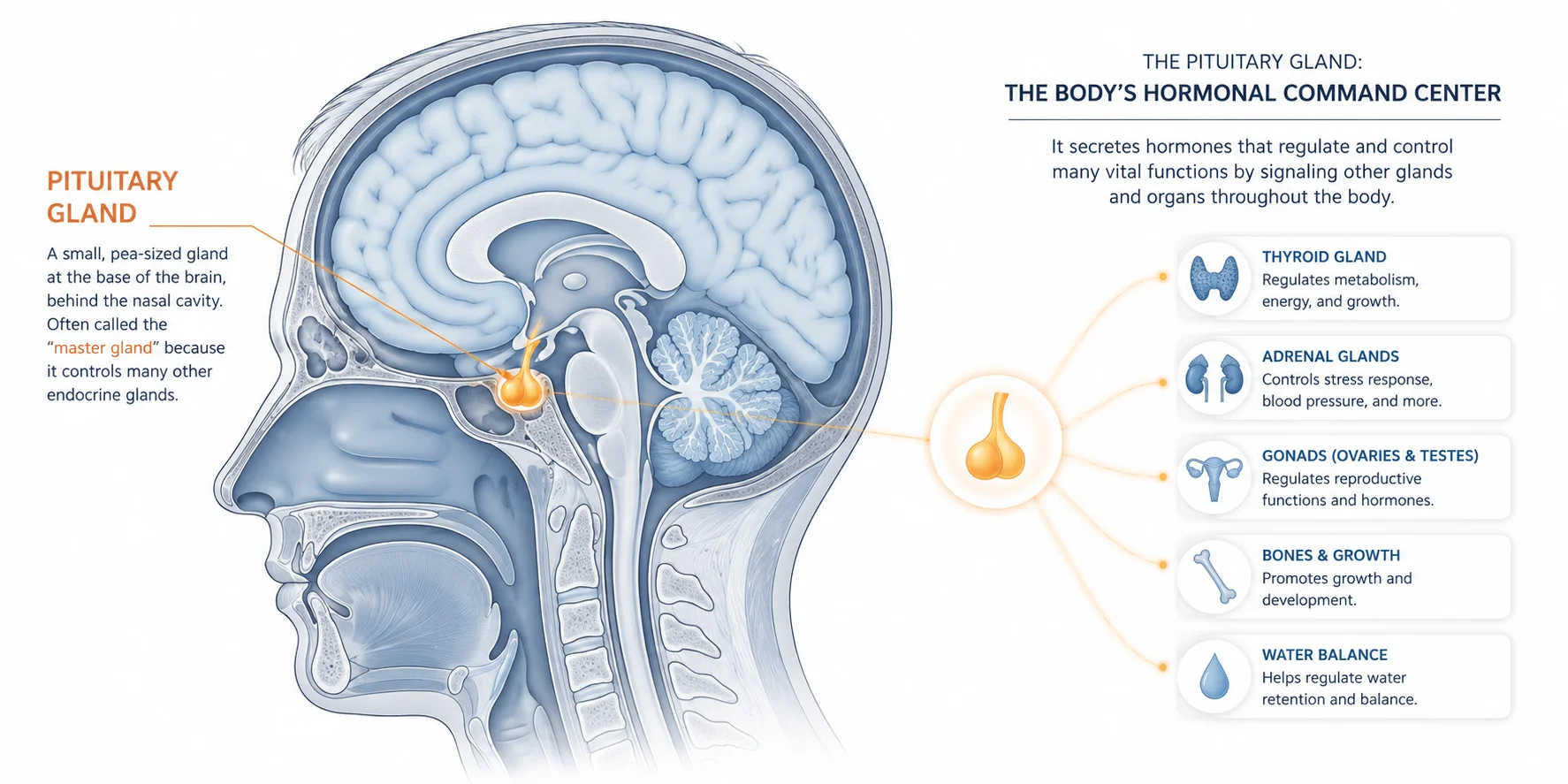
Hearing the word “hypophysectomy” from a doctor usually means one thing has already happened: something showed up on an MRI. This is surgery to remove the pituitary gland, in part or in full, and it’s almost never a first-line treatment. It’s what comes after other options have been tried, or after a scan reveals a growth that’s too risky to leave alone.
This overview covers what the procedure actually involves, who tends to need it, how it’s typically performed today, and what recovery realistically looks like. It isn’t a substitute for a conversation with a neurosurgeon or endocrinologist. Every case here comes down to gland anatomy, tumor size, and personal health history, which are exactly the details a specialist reviews before recommending surgery.
What the Pituitary Gland Does, and Why Removing It Is a Big Deal
The pituitary is small, roughly the size of a pea, sitting at the base of the brain just behind the bridge of the nose. Despite its size, it functions as the control center for the body’s hormone system. It signals the thyroid, adrenal glands, and reproductive organs, and it directly produces hormones tied to growth, metabolism, and stress response.
That’s why removing it isn’t a decision made lightly. Take away the gland, and the body loses its main hormonal messenger. Patients typically need lifelong hormone replacement afterward, replacing what the gland used to produce on its own. The tradeoff, though, is that hormone replacement is well understood and generally manageable. A tumor left growing near the brain and optic nerves is a much less predictable risk.
Why Hypophysectomy Gets Recommended
The most common reason is a pituitary adenoma, a tumor on the gland itself. Surgery becomes the recommended path when a nonsecreting adenoma is causing symptoms, especially visual problems like peripheral vision loss, or when imaging shows it’s continuing to grow. Some adenomas also secrete excess hormones, and shrinking or removing the tumor is the most direct way to bring those levels back down.
Cushing’s syndrome is another common trigger. This condition comes from the body’s tissues being exposed to too much cortisol for too long, usually because of a benign pituitary adenoma, and surgery aims to remove the tumor while preserving as much of the healthy gland as possible.
Less commonly, hypophysectomy is used for craniopharyngiomas, a rare tumor type, or pituitary carcinoma. There’s also a narrower historical use worth mentioning: the procedure has been used to relieve severe, opioid-resistant cancer pain in patients whose pain couldn’t be controlled through standard interventional treatments. This isn’t a common indication today, but it shows up in the medical literature and occasionally in patient questions, so it’s worth knowing it exists.
How the Surgery Is Actually Done
There are three general techniques, and which one a surgical team picks depends on tumor size, location, and how much of the gland needs to come out.
Transsphenoidal surgery is by far the most common approach now. Instead of opening the skull, the surgeon reaches the gland through the nose and sphenoid sinus, working with an endoscope and a set of fine instruments. The path goes in beneath the upper lip and into the nasal cavity, where a speculum and specialized forceps are used to access and remove the gland or tumor. No external incision, no visible scarring, and generally a faster recovery than older approaches.
This method has an interesting history. The first transsphenoidal pituitary surgery was performed in 1907 by an Austrian surgeon named Hermann Schloffer, using a three-stage operation that required removing part of the sphenoid and ethmoid sinuses just to reach the gland. The original patient developed a serious complication and didn’t survive long after surgery. It took decades of refinement, better imaging, and eventually endoscopic tools, to turn that risky first attempt into the routine, minimally invasive procedure it is now.
Open craniotomy is the older, more invasive option, reserved for tumors that are too large, awkwardly positioned, or otherwise unreachable through the nose. It involves opening the skull directly.
Stereotactic radiosurgery isn’t traditional surgery at all. It uses tightly focused radiation to target and destroy pituitary tissue without any incision. It’s typically considered when a patient isn’t a good candidate for either surgical approach, or as a follow-up if some tumor remains after an initial operation.
Before any of these happens, a fairly standard workup takes place: hormone level testing, an MRI to map the tumor precisely, and a pre-surgical evaluation covering blood work and heart health to confirm the patient can safely handle anesthesia.
What Recovery Looks Like
Transsphenoidal surgery typically runs about two hours. Afterward, the nose is packed to control bleeding, and most patients go home the next day, which is a notably shorter hospital stay than open craniotomy requires.
A few complications are worth knowing about going in, not because they’re likely, but because catching them early matters:
- Diabetes insipidus. This shows up as unusual thirst and frequent urination in the first few days after surgery. Most borderline cases resolve on their own within a few days as the back part of the pituitary regains function. A small number of patients need ongoing treatment for it.
- CSF leakage. Because the gland sits so close to the brain and its surrounding fluid, a cerebrospinal fluid leak is possible. If it happens during the operation, the surgical team repairs it right away; if it happens later, after the nasal packing comes out, it may need additional treatment.
- Infection. This is a serious but uncommon risk, and the vast majority of cases respond well to antibiotics, which patients are typically given during surgery and until the nasal packing is removed.
- Hormonal changes. Imbalances can persist or worsen after the operation, which is why regular follow-up with the surgical and endocrine team matters just as much as the surgery itself.
If the gland is removed entirely, hormone replacement becomes a permanent part of life afterward. Cortisol replacement usually comes first and matters most, since the adrenal system depends on it. Thyroid hormone and other replacements follow depending on which functions were lost. None of this is exotic medicine; it’s a well-established, manageable part of care that an endocrinologist monitors long-term, adjusting doses based on regular bloodwork.