
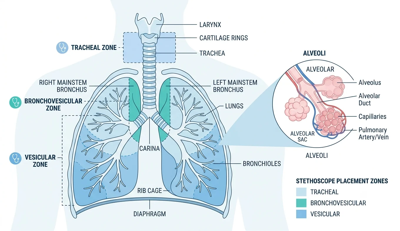
Place a stethoscope on almost any point across a healthy person’s back or chest, and you’ll hear the same thing: a soft, low rustle that swells on the inhale and fades almost as soon as it starts on the way out. That sound is the vesicular breath sound, and it’s the baseline every other lung finding gets measured against.
It’s easy to skip past vesicular sounds in training because they’re “just normal.” But normal is exactly the point. You can’t recognize a diminished breath sound, a consolidation, or a pneumothorax if you don’t have a clear reference for what healthy air movement actually sounds like. This guide covers what vesicular breath sounds are, where they come from, how they compare to bronchial and bronchovesicular sounds, and what it means clinically when they change.
What Are Vesicular Breath Sounds?
Vesicular breath sounds are the soft, low-pitched breath sounds heard over most of the lung surface during normal breathing. They are soft, low-pitched, predominantly inspiratory, and heard especially well at the posterior lung bases. The inspiratory phase is louder and lasts longer than expiration, which fades quickly, sometimes described as an inspiration-to-expiration ratio of roughly 3:1, though some sources put it closer to 2:1.
The name comes from where the sound is generated. Vesicular sounds normally arise from the finer lung parenchyma, meaning the small airways and alveoli out at the lung periphery, rather than the large central airways. That’s a useful anchor point: bronchial sounds start centrally and are abnormal if heard peripherally; vesicular sounds start peripherally and are abnormal if heard where bronchial sounds should be.
One clarification worth making explicit: despite the name, vesicular breath sounds aren’t produced by air moving in the alveoli themselves. The inspiratory component of vesicular breath sounds originates in the peripheral portions of the lung near where the stethoscope is placed, generated by turbulent airflow in the smaller bronchi and bronchioles, then filtered and softened by the surrounding lung tissue before it reaches the chest wall.
How Vesicular Sounds Compare to Other Breath Sounds
Clinicians typically describe three categories of normal breath sounds, and the differences come down to where you’re listening and what’s underneath the stethoscope.
| Feature | Vesicular | Bronchovesicular | Bronchial |
|---|---|---|---|
| Pitch | Low | Medium | High |
| Intensity | Soft | Moderate | Loud, harsh |
| Inspiration vs. expiration | Inspiration longer and louder | Roughly equal | Expiration longer and louder |
| Normal location | Most of the lung surface, especially posterior bases | Upper anterior chest, between the scapulae | Over the trachea, right upper sternal border |
| Origin | Small airways and alveoli at the lung periphery | Transition zone between large and small airways | Large central airways (trachea, mainstem bronchi) |
Bronchial sounds, also called tubular sounds, normally arise from the tracheobronchial tree, while vesicular sounds normally arise from the finer lung parenchyma. Bronchovesicular sounds can be heard during both inspiration and expiration and have a mid-range pitch and intensity, commonly over the upper third of the anterior chest.
The clinical trap here isn’t identifying vesicular sounds where you expect them. It’s catching bronchial sounds outside their normal territory, since that combination usually signals consolidation, and recognizing when vesicular sounds are unexpectedly quiet or missing.
When Vesicular Breath Sounds Are Diminished or Absent
A reduction in vesicular breath sounds is one of the more common, and more useful, abnormal findings in a respiratory exam. Clinicians often grade intensity on a simple scale: zero for absent, one for barely appreciated, two for diminished, three for normal, and four for louder than normal, though the practical value of this grading has been debated.
A few mechanisms explain why the sound drops off:
Reduced airflow. In obstructive disease, less air is moving through the small airways to begin with, so there’s simply less sound to transmit. This shows up in asthma and COPD, where diminished sounds often travel alongside prolonged expiration or wheezing.
Something blocking transmission. Air is a poor conductor of sound compared to fluid, so anything that inserts air, fluid, or tissue between the alveoli and the stethoscope will muffle or erase the sound. If fluid, air, or other matter lies in the pleural space, it will decrease the intensity, as happens with a pleural effusion. A pneumothorax works the same way from the opposite direction: air trapped in the pleural space, rather than fluid, still blocks transmission.
Loss of functioning lung tissue. Conditions like emphysema destroy alveolar architecture, so there’s less tissue capable of generating and transmitting the sound in the first place.
One finding is worth flagging on its own: asymmetry. An asymmetric intensity of breath sounds is especially helpful after intubation, where asymmetric breath sounds are pathognomonic for accidental placement of the tube into a single bronchus rather than the trachea. If one side of the chest is clearly quieter than the other on an otherwise identical exam, that’s a finding to chase down, not dismiss as normal variation.
Why This Matters Beyond the Exam Room
Vesicular breath sounds don’t diagnose anything by themselves. Their value is as a baseline. Once you know what normal sounds like at a given location, a change from that baseline, whether it’s reduced intensity, a shift toward bronchial quality, or new asymmetry, becomes a genuine clinical signal rather than background noise.
That’s also why auscultation technique matters as much as recognition. A quiet room, direct skin contact rather than through clothing, and a full inhale-exhale cycle at each site all affect whether a soft sound is genuinely diminished or just poorly heard. Comparing side to side at matching locations, rather than just moving down one side of the chest, is what actually surfaces asymmetry.