Hypoxemic respiratory failure happens when the lungs can no longer move enough oxygen into the blood, even though the body is still able to get rid of carbon dioxide. Clinicians define it by a specific number: an arterial oxygen level, or PaO2, below 60 mmHg. That threshold isn’t arbitrary. Below it, hemoglobin’s ability to hold onto oxygen drops off sharply, and organs, especially the brain and heart, start running short on the fuel they need to function.
This is also called type I respiratory failure, to separate it from type II (hypercapnic) failure, where the main problem is a buildup of carbon dioxide rather than a shortage of oxygen. The distinction matters because the two types come from different mechanical problems in the lungs, and they’re often treated differently.
What Actually Goes Wrong in the Lungs
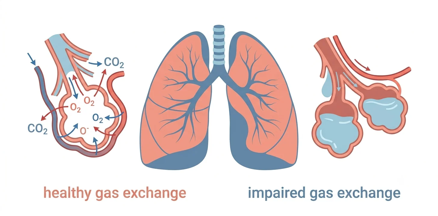
Healthy gas exchange depends on air (ventilation) and blood flow (perfusion) meeting up properly in the tiny air sacs of the lungs, called alveoli. Hypoxemic respiratory failure develops when that meeting breaks down, through one of a few mechanisms:
- Ventilation-perfusion (V/Q) mismatch. Blood flows past alveoli that aren’t getting enough air, or air reaches alveoli with poor blood flow. This is the most common cause of hypoxemia overall.
- Shunt. Blood passes through the lungs without ever reaching a functioning alveolus, so it picks up no oxygen at all. This happens when alveoli are filled with fluid or collapsed, as in pneumonia or pulmonary edema, and it’s notable because supplemental oxygen alone often doesn’t fix it.
- Diffusion limitation. The barrier between the alveolus and the blood vessel becomes too thickened or damaged for oxygen to cross efficiently, seen in conditions like severe fibrosis.
- Low inspired oxygen. Rare outside of high altitude or unusual environments, this simply means there isn’t enough oxygen in the air being breathed in.
Acute hypoxemic respiratory failure is defined by a PaO2 below 60 mmHg or an SaO2 below 88%, and it may result from V/Q mismatch, shunt, hypoventilation, diffusion limitation, or low inspired oxygen tension.
What Causes It
Hypoxemic respiratory failure isn’t a disease on its own. It’s the end result of something else going wrong in the lungs or heart. It’s typically caused by intrapulmonary shunting of blood with resulting V/Q mismatch, driven by airspace filling or collapse from cardiogenic or non-cardiogenic pulmonary edema, pneumonia, or pulmonary hemorrhage, by airway disease such as asthma or COPD, or by intracardiac shunting of blood from the right side of the heart to the left.
The single biggest driver is acute respiratory distress syndrome, or ARDS, a severe inflammatory lung injury. ARDS is a type of acute hypoxemic respiratory failure caused by diffuse inflammatory lung injury, and it accounts for roughly two-thirds of all acute hypoxemic respiratory failure cases. Sepsis and pneumonia together cause the majority of ARDS cases. Other common triggers include:
- Severe bacterial, viral, or fungal pneumonia
- Cardiogenic pulmonary edema, where a failing heart backs fluid up into the lungs
- Pulmonary embolism, a blood clot blocking flow through part of the lung
- Aspiration of stomach contents or other material into the airway
- Trauma, severe pancreatitis, or major surgery, all of which can trigger ARDS indirectly through systemic inflammation
Age and existing health conditions raise the stakes. People with chronic lung disease, heart failure, or a weakened immune system tend to decompensate faster and have a harder time recovering.
Recognizing the Warning Signs
The symptoms of hypoxemic respiratory failure track the underlying oxygen shortage, and they tend to escalate as PaO2 keeps falling. Common symptoms include shortness of breath, rapid breathing, extreme tiredness, a fast heart rate, and in some cases coughing up blood. As oxygen levels drop further, more serious signs appear.
Low blood oxygen causes a bluish coloration of the skin in people with lighter skin tones, and a gray or whitish coloration in the mouth, around the eyes, and under the nails in people with darker skin tones. This is called cyanosis, and it’s one of the more visible signs that oxygen delivery has dropped to a dangerous level. Confusion, drowsiness, and agitation often follow, since the brain is especially sensitive to falling oxygen. Left uncorrected, the brain and heart begin to malfunction, leading to unconsciousness and abnormal heart rhythms.
It’s worth knowing that these signs aren’t perfectly reliable on their own. A fast heart rate, rapid breathing, and mental status changes are non-specific and can occur for reasons unrelated to low oxygen, and some people who are chronically hypoxemic show no obvious physical signs at rest. That’s exactly why clinicians rely on direct measurement rather than symptoms alone.
Anyone showing sudden shortness of breath together with bluish or gray skin discoloration, confusion, or extreme fatigue needs emergency medical attention right away. This combination can point to a rapidly worsening oxygen shortage, and it’s not something to wait out at home.
How It’s Diagnosed
Diagnosis starts with a pulse oximeter, the small clip-on sensor placed on a fingertip that estimates blood oxygen saturation without needles. If that reading is low or the clinical picture is concerning, an arterial blood gas test follows. This involves drawing blood directly from an artery, usually at the wrist, to get an exact PaO2 value along with carbon dioxide and pH levels.
Clinicians also look at the ratio of PaO2 to the fraction of inspired oxygen a person is receiving, written as PaO2/FiO2 or the “P/F ratio.” A PaO2/FiO2 ratio under 300 is one of the defining features used in the Berlin criteria for ARDS, alongside acute onset, bilateral infiltrates visible on a chest X-ray, and the absence of heart failure as the primary cause. A chest X-ray or CT scan typically follows to help pin down the underlying cause, whether that’s fluid, infection, collapse, or something else.
How Hypoxemic Respiratory Failure Is Treated
Treatment follows a step-up approach, starting with the least invasive option that gets oxygen levels back to a safe range and escalating only as needed.
Supplemental oxygen through a nasal cannula or face mask is usually the first step for milder cases. High-flow nasal cannula (HFNC) delivers warmed, humidified oxygen at much higher flow rates and has become a mainstay for more significant hypoxemia. Non-invasive ventilation, including CPAP and BiPAP delivered through a tight-fitting mask, provides breathing support without a tube in the airway. When these aren’t enough, invasive mechanical ventilation through a breathing tube takes over the work of breathing entirely, buying time for the underlying cause to be treated.
Guideline-based management of acute respiratory failure emphasizes restrictive fluid strategies in patients without shock, and high-flow nasal cannula oxygenation is now weakly recommended for general respiratory support and for initial management of ARDS. Alongside respiratory support, treatment always targets the root cause, whether that’s antibiotics for pneumonia, diuretics for fluid overload, or anticoagulation for a blood clot.
Outcomes vary widely depending on the cause and how quickly treatment starts. ARDS carries a substantial mortality risk, with a 28-day mortality rate of roughly 35% and an overall in-hospital mortality rate of around 40% in some cohorts. Milder cases caught early and treated with oxygen alone generally have a far better outlook.