
A patient comes in three days after starting a new gym program, complaining of dark urine and thighs so sore he can barely climb stairs. Or it’s a trauma patient pulled from a car wreck. Or an elderly woman found down on her kitchen floor after a fall, unable to get up for sixteen hours. Different stories, same diagnosis to rule out: rhabdomyolysis.
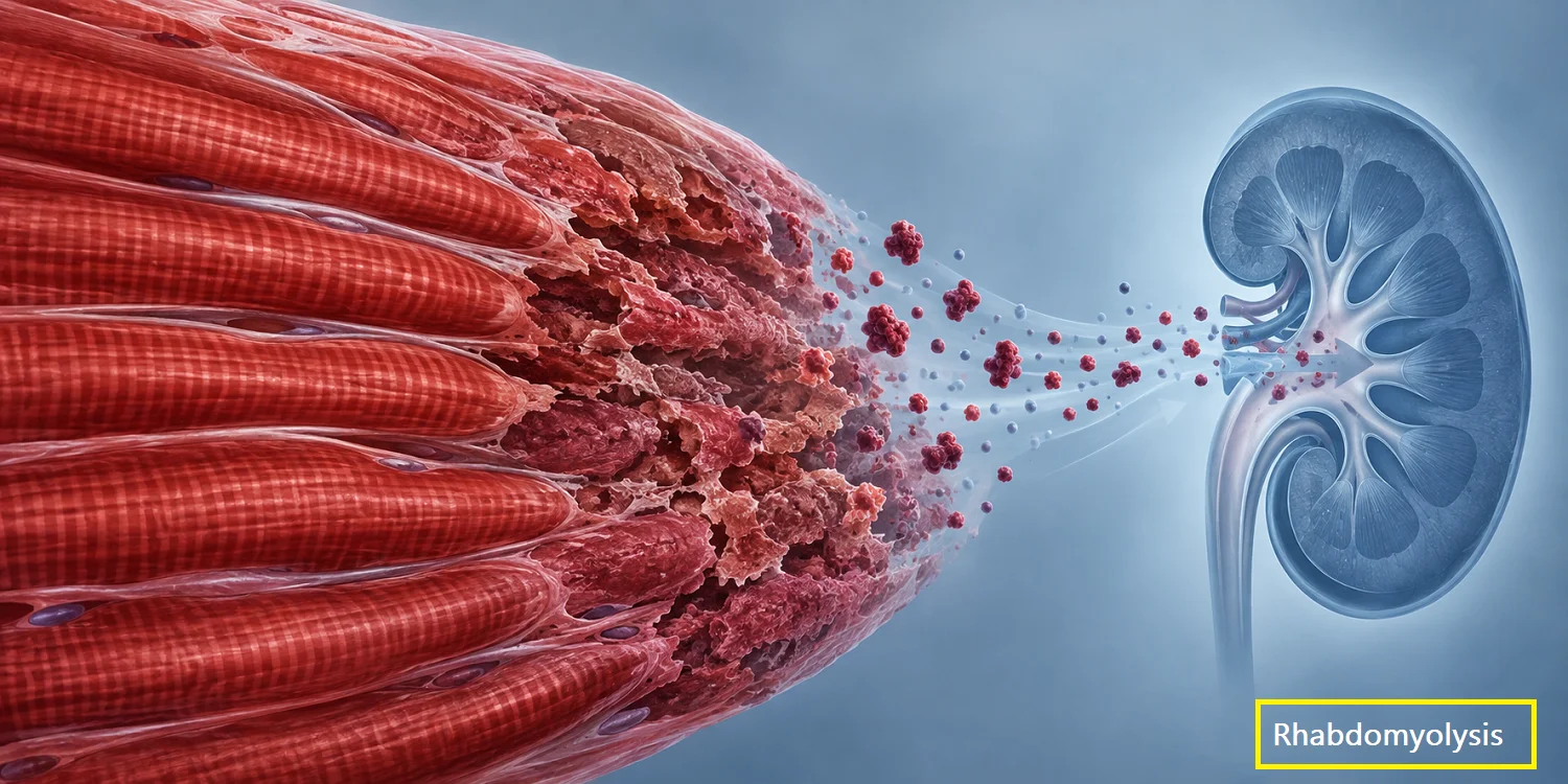
Rhabdomyolysis is the breakdown of skeletal muscle with release of intracellular contents, chiefly creatine kinase (CK), myoglobin, and potassium, into the bloodstream. It’s not a single disease but a final common pathway that dozens of different insults can lead to. The clinical spectrum runs from an asymptomatic CK bump discovered incidentally on labs to a patient in acute kidney injury with a potassium high enough to stop their heart. That range is exactly why it’s easy to underestimate at the bedside.
What Counts as Rhabdomyolysis, Exactly?
For a long time, “rhabdomyolysis” was one of those diagnoses everyone recognized but nobody had fully agreed how to define. A 2024 international workshop (the 276th ENMC workshop, bringing together 21 specialists) tried to fix that. The group agreed that a diagnosis of rhabdomyolysis requires clinical symptoms of severe muscle swelling, weakness, or myalgia, plus serum CK levels exceeding 10,000 IU/L for exertional cases and above 5,000 IU/L for non-exertional cases, with CK peaking one to four days after the triggering event and returning to baseline within one to two weeks of rest. [1]
That’s a stricter bar than what most clinicians grew up using. The older and still widely cited rule of thumb is simpler: a CK rise to five times the upper limit of normal or greater, following intense physical activity or injury, alongside the classic triad of myalgia, weakness, and myoglobinuria. [2] In practical terms, since normal CK sits roughly in the 30 to 200 IU/L range depending on the lab, a level above about 950 to 1,000 IU/L is generally treated as diagnostic. [3]
Why does the exact cutoff matter? Because it changes who gets labeled “at risk of AKI” and who gets aggressive fluids versus outpatient follow-up. A CK of 1,200 in an otherwise well patient after a hard leg day at the gym is a very different clinical problem than a CK of 40,000 in a found-down patient. Use the number, but don’t let it replace judgment.
One more diagnostic quirk worth knowing: urine dipstick will read positive for blood in myoglobinuria even though there are no red cells on microscopy. That mismatch, blood-positive dipstick with a clean urine microscopy, is a fast bedside clue that you’re dealing with pigment, not hematuria.
Why Muscle Breakdown Becomes a Kidney Problem
The pathophysiology is worth walking through because it explains every downstream decision. When muscle fibers die, they dump myoglobin, potassium, phosphate, and various intracellular enzymes into the circulation. Myoglobin gets filtered by the kidney and can precipitate in the renal tubules, especially in an acidic, volume-depleted environment. That precipitation, combined with direct heme-mediated oxidative injury to tubular cells, is the main mechanism behind rhabdomyolysis-induced acute kidney injury.
This is also why the two pillars of early management, volume and urine pH, aren’t arbitrary. Adequate hydration keeps urine flowing and dilutes the myoglobin load. Alkalinizing the urine (when used) is meant to reduce the precipitation of myoglobin casts. Neither intervention fixes the muscle damage that’s already occurred; both are aimed squarely at protecting the kidney from what the muscle damage is dumping into it.
Who Gets Rhabdomyolysis, and From What
The causes fall into a few broad buckets, and most cases have more than one contributing factor stacked together, a patient on a statin who then runs a marathon in the heat, say.
Trauma and physical causes. Crush injuries, prolonged immobilization (a fall with a long “found down” interval, surgical positioning, unconsciousness from alcohol or overdose), burns, and electrical injury all cause direct mechanical or ischemic muscle damage.
Exertional causes. Marathon runners, novice gym-goers doing high-intensity eccentric exercise, military recruits, and athletes training in heat are classic populations. This group tends to skew toward the higher CK thresholds in the newer diagnostic criteria, since exercise-induced CK elevations are common and only the more extreme rises should be labeled pathological.
Drugs and toxins. Statins cause direct myotoxicity, and the list of other drugs implicated includes cyclosporine, colchicine, and corticosteroids, while alcohol, cocaine, and amphetamines cause indirect muscle damage. [4] Statin-associated rhabdomyolysis is rare in absolute terms, generally cited at well under 1% of statin users, but it’s common enough as a differential consideration that it belongs on every medication reconciliation for a patient presenting with myalgia and CK elevation.
Infections. Influenza, HIV, and various bacterial infections can trigger rhabdomyolysis, and in pediatric populations infection may account for a notably larger share of cases than it does in adults.
Metabolic and inherited causes. Severe electrolyte derangements (hyponatremia, hypokalemia), inherited metabolic myopathies, and malignant hyperthermia round out the list. These are the cases that should raise suspicion when a patient has recurrent episodes, a family history, or an episode triggered by minimal exertion, that pattern is a signal to think about genetic testing rather than assuming another one-off exertional event.
The Workup
A patient with suspected rhabdomyolysis needs a focused but specific set of labs, not a shotgun panel.
Creatine kinase, drawn serially rather than once, since CK typically rises within two to twelve hours of muscle injury and stays elevated for days [2], so a single normal value early in the course doesn’t rule it out. Peak CK, not the admission value, correlates better with outcomes like need for renal replacement therapy.
Renal function and electrolytes. Creatinine, potassium, calcium, and phosphate all need close attention. Hyperkalemia from cell lysis is the acute life-threatening complication to watch for, arrhythmia can happen fast and is arguably the most immediately dangerous part of the whole syndrome.
Urinalysis, specifically looking for that blood-positive, RBC-negative pattern described above.
Additional testing is often unnecessary. Imaging, muscle biopsy, or electromyography are not required to diagnose rhabdomyolysis [5] in the acute setting; biopsy has a role later, after recovery, when an underlying inflammatory or metabolic myopathy is suspected because of recurrence or an atypical trigger.
Also worth building into the workup: a coagulation panel in more severe cases, since disseminated intravascular coagulation is a recognized, if less common, complication.
Managing the Patient in Front of You
The first step is identifying and removing the underlying cause where possible [5], stopping the offending statin, addressing the compartment syndrome, correcting the electrolyte disturbance driving it. Everything after that is supportive.
Fluids are the cornerstone. Preservation of renal function with intravenous fluid therapy remains the backbone of rhabdomyolysis treatment [6], and the evidence for this goes back decades: in a group of disaster patients treated with on-site IV fluids in 1984, none went on to develop acute kidney injury. [6] Aggressive early hydration, on the order of 1.5 liters per hour in some protocols [7], aiming for a urine output goal around 200 mL/hour, is the standard approach in moderate to severe cases. Some protocols alternate normal saline with a bicarbonate-containing solution to alkalinize the urine, though this remains more variable in practice than the fluid resuscitation itself.
There’s less consensus than you’d expect. No formal treatment guidelines exist for rhabdomyolysis, and no randomized controlled trials have directly tested its management [6], which is a little startling for a condition this common and this well described. Most of what clinicians do is built on physiological reasoning, retrospective cohorts, and long clinical experience rather than trial data. Worth knowing when a patient or family asks why the “protocol” seems to vary between providers.
Watch for the downstream complications, not just the kidney. Beyond AKI, keep an eye on:
- Compartment syndrome, particularly in crush injury or prolonged-immobilization cases; this needs surgical evaluation, not just monitoring
- Hyperkalemia, checked serially, with a low threshold for cardiac monitoring
- Disseminated intravascular coagulation in more severe presentations
- Cardiac arrhythmia related to electrolyte shifts
Risk-stratify rather than treating every elevated CK the same way. The McMahon score, a prediction model built from admission demographics, clinical variables, and labs, is one tool used to estimate which patients are genuinely at risk for renal failure and need the aggressive protocol versus who can be managed more conservatively. In one retrospective study of 232 rhabdomyolysis patients, 19% developed AKI and 12.5% ultimately required renal replacement therapy [8], so the majority of patients do recover without needing dialysis, but the minority who don’t tend to do so quickly, which is the whole argument for early aggressive management rather than a wait-and-see approach.
Outcomes and What to Tell Patients
Prognosis depends heavily on whether AKI develops. Mortality without AKI is around 20%, rising to as much as 50% when AKI occurs [9], figures that reflect sicker overall patient populations (trauma, sepsis, prolonged downtime) as much as the rhabdomyolysis itself, so they shouldn’t be quoted to a healthy 24-year-old with post-workout myalgia and a CK of 3,000 without that context. Most exertional and mild drug-induced cases resolve fully with hydration and rest, no long-term sequelae, no residual kidney damage.
Patients who’ve had one episode, especially without an obvious trigger like a single hard workout, deserve a conversation about recurrence risk and, in the right clinical picture, referral for a metabolic or neuromuscular workup. The 2024 ENMC consensus specifically flagged follow-up, including physical and psychological effects, as an area that’s been under-addressed relative to the acute management literature.
Conclusion
Rhabdomyolysis rewards a structured approach: confirm the diagnosis with CK trends rather than a single number, identify and remove the trigger, hydrate aggressively while monitoring for hyperkalemia and compartment syndrome, and risk-stratify for AKI rather than treating every case identically. The evidence base is thinner than the condition’s familiarity would suggest, so clinical judgment still carries real weight here, particularly in borderline CK elevations where the newer, stricter diagnostic thresholds and the older five-times-normal rule can point in different directions.